If you don’t know the difference between an MRI and a CT scan, you are a member of a large club. Both imaging tests involve large machines which surround or enclose your body; both are very expensive but usually paid for by health insurance; and both provide pictures of what’s going on inside your body to help doctors pinpoint the source of your problem. Either test may be administered with an injected dye to highlight certain areas.
If you come to one of the offices of Long Island Spine Rehabilitation Medicine seeking pain relief and improved range of motion for your back or joints, you have come to the right place; our doctors specialize in nonsurgical treatments of spinal and musculoskeletal disorders. Depending on your symptoms, your medical history, and the results of our physical examination, we may recommend that you undergo further testing, often including either an MRI or CT scan.
This article is designed to clarify the nature of both exams and to explain why we may choose one over the other in your particular case. It is also intended to prepare you for what each test feels like and to relieve any anxiety you may have about having one administered.
How does a CT scan produce images?
Computed tomography (CT or CAT scan) uses the same technology as an X-ray machine (ionizing radiation), but the CT scan is linked to a computer. Because the beams of the CT scan device rotate around your body as you lie on a table, the images taken are cross-sectional slices that provide far more insight into what’s going on than a single image ever could.
How does an MRI scan produce images?
The name “MRI” is an acronym for magnetic resonance imaging. An MRI device uses a combination of a strong magnet, a radio transmitter, and a receiver. While electric current is sent through a coiled electromagnet, the switching of the currents causes the coils to expand. This process results in the loud clicking sounds you will hear during the procedure.
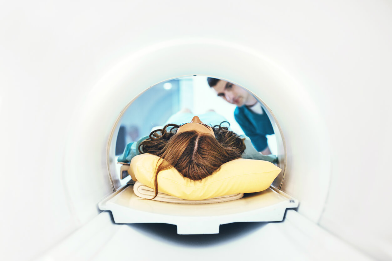
The resulting images of your body’s internal structures are created by the interaction between the powerful magnetic, the radio frequency pulses, and the computer. During an MRI, you typically lie on a flat table that slides into a tube or tunnel for the duration of the test.
What are the advantages of each imaging technique?
Simply put, it depends on whether your doctor is examining skeletal or soft tissue. Generally speaking, CT scans provide better visuals of your bones and blood vessels, while MRI scans show more defined pictures of your organs, muscles, tendons, ligaments, and intervertebral discs.
Both imaging tests can be read and interpreted by a diagnostic radiologist immediately after the test, but it usually takes a day or two (unless there is an urgent need) for you to hear the results. The CT test typically takes 5 to10 minutes, while the MRI takes 30 to 60 minutes. For both tests, when the machinery is actively taking pictures, you have to remain still.
What are the potential drawbacks of each test?
Both MRI and CT scans are considered safe, with a small number of exceptions. Some patients may be allergic to dye injected for either test, so healthcare providers are always prepared to deal with serious allergic reactions.
Because CT scans use radiation, some patients who have been exposed to extensive radiation previously may want to avoid these tests, although the exposure is brief. Because the ionizing radiation used in CT scans has an association with cancer, doctors don’t administer CT scans when they are not absolutely necessary. Still, the risk of a CT scan leading to cancer is extremely small.
MRI scans, on the other hand, are not a viable option for all patients. If you have certain types of metal in your body, such as a pacemaker, cochlear implant, insulin pump, or spinal cord stimulator, having an MRI may be dangerous. The metal used in most orthopedic plates and heart stents, however, has been ruled to be entirely safe by the National Institutes of Health.
Nonetheless, MRI scans are problematic for some patients for other reasons. Some people find the loud repetitive noises disturbing (even when muffled by the earmuffs provided), and others become claustrophobic being enclosed inside the tunnel of the machine. Fortunately, in recent years, open MRIs have become available, both to make claustrophobic patients more comfortable and to accommodate patients whose size or weight makes traditional MRI equipment impractical.
When you visit our offices, you can be sure our doctors will discuss all diagnostic and treatment options with you before making any decisions.