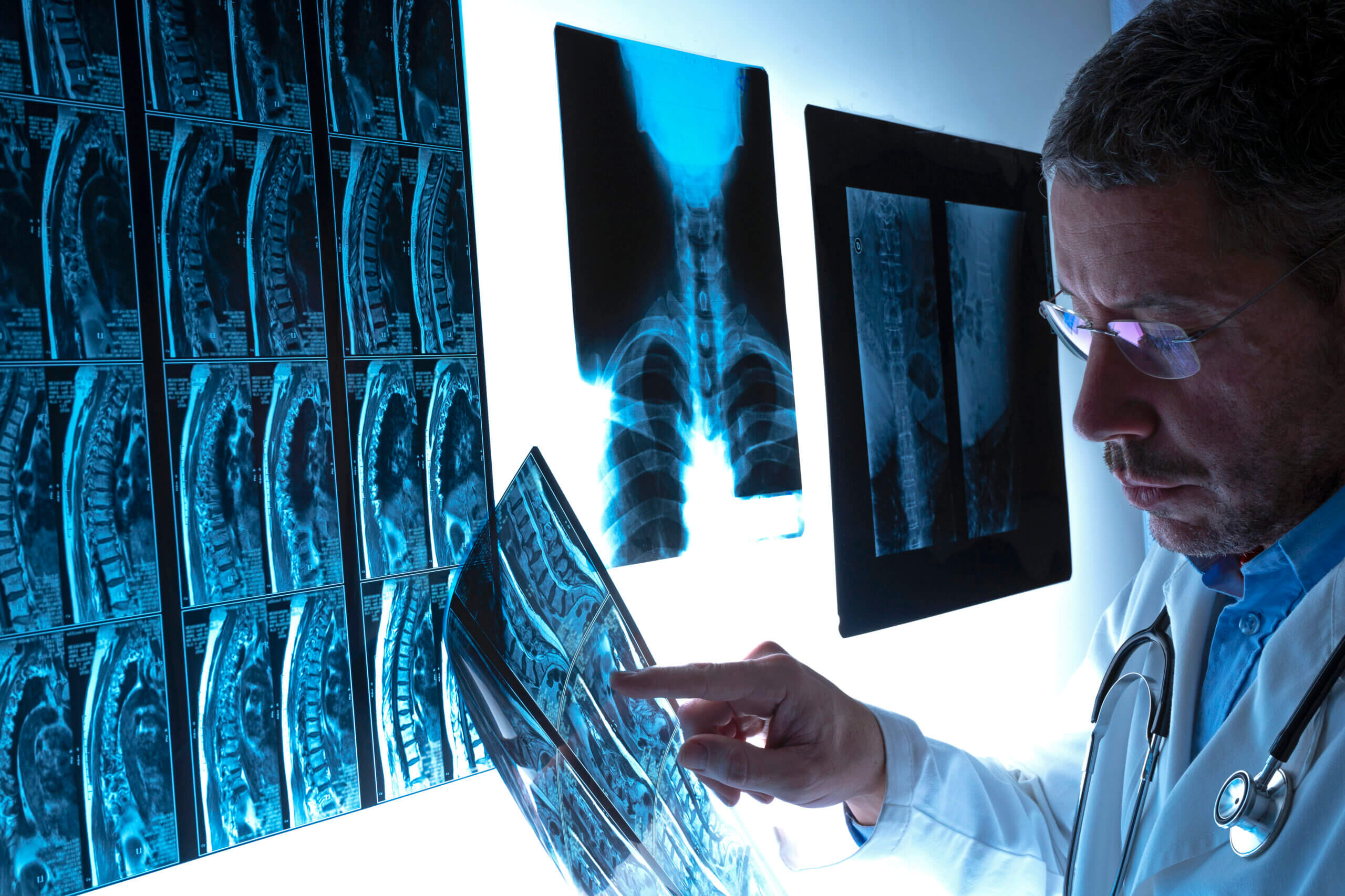
Gone are the days when X-rays were the only diagnostic imaging test. Now, when you are experiencing musculoskeletal pain or impaired mobility and you seek medical treatment, your doctor has several possible diagnostic imaging methods readily available.
This is especially true at Long Island Spine Rehabilitation Medicine where our talented physiatrists have access not only to cutting-edge imaging tests, but also to electromyography (EMG), nerve conduction studies, and diagnostic injections. Let’s take a look at the types of diagnostic imaging methods our doctors use and how they differ from one another:
X-Rays
X-rays can penetrate most objects and so can give a clear view of the inside of the human body. When you go to the doctor presenting with severe joint or back pain, X-rays can, within 10 to 15 minutes, provide several images of the bones of the affected region.
Nonetheless, the electromagnetic radiation of the X-rays may be harmful, especially if administered to an unborn child, and over years have a cumulative effect. This is why lead aprons are used to protect patients who receive X-rays. Despite this drawback, however, X-rays are accurate and the least expensive of the imaging tests.
CT Scans
A computerized tomography or CT scan, records X-ray images taken from several vantage points, using computer processing to produce cross-section pictures of bone, blood vessels, and some soft tissues. They can show fractures, joints eroded by arthritis, bone tumors, and even blood clots. Because the images are taken at various angles, CT scans provide more detailed images than individual X-rays.
Although CT scans, like X-rays, also use potentially damaging radiation, they have the advantage of providing detailed, rapid results. When the situation is urgent and/or the patient is in extreme pain, the fact that CT scan images can be created within 10 to 15 minutes can be of great benefit.
MRI Scans
MRI scans have a major advantage when it comes to diagnostic imaging tests in that they provide much clearer pictures of soft tissue injuries and abnormalities, as well as images of bone. With an MRI scan, our doctors can examine joints, cartilage, tendons, bone fractures, and peripheral nerves with much greater clarity. MRI scans also show evidence of inflammation and infection.
There are, however, disadvantages to MRIs, including that the equipment involved is narrower, deeper, and more enclosed than that used for CT scans. As a result, patients may become claustrophobic during MRIs, though there are open MRIs available at some venues. MRIs also take longer than other imaging tests, lasting an average of 45 minutes to 1 hour, and are significantly more expensive than the other tests mentioned
Ultrasound
Ultrasound tests, administered manually with a transducer (something like the mouse on a computer), provide the benefit of dynamic images that show moving images in real-time. They are also cost-effective, relatively quick (20 to 40 minutes), and completely safe since they do involve ionizing radiation.
Ideal for imaging superficial musculoskeletal structures, ultrasound successfully uses sound waves to produce pictures of muscles, tendons, ligaments, nerves, and joints throughout the body. At Long Island Spine Rehabilitation Medicine, we use it to help diagnose sprains, strains, tears, trapped nerves, arthritis, and other musculoskeletal conditions.
Contact Our Experienced Doctors Today
At Long Island Spine Rehabilitation Medicine, we know the importance of accurate diagnosis. Once we diagnose the cause of your problem, we will resolve it with a treatment program designed especially for you. The sooner you contact us, the sooner you will start feeling better.
